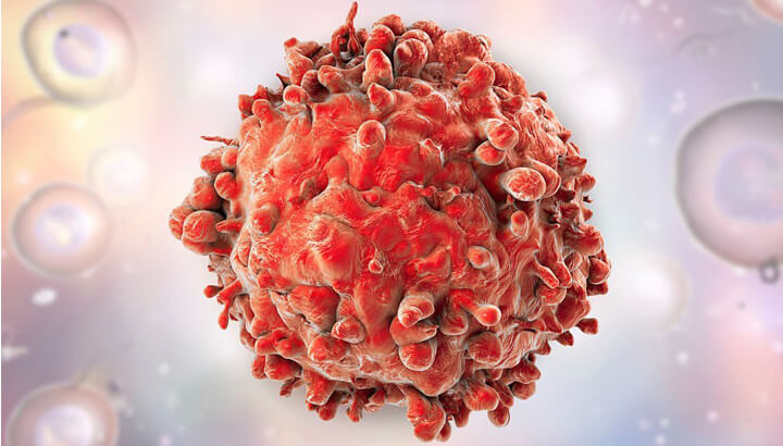
Leukemia is a broad term for all types of cancers of the blood cells, including the cancers of the bone marrow cells and the lymphatic system. According to Globocan 2018, the worldwide incidence rate of leukemia in 2018 was 3.2 per 100,000 population. In India, about 42,055 new cases of leukemia were reported in 2018.
Acute leukemia refers to the rapidly progressing cancer that originates in the blood-forming tissues. This cancer causes the production of a large number of abnormal white blood cells; these cells are immature and non-functional and crowd out the mature, functional cells. Based on the type of white blood cells affected, acute leukemia can be classified as acute myelocytic leukemia (AML) and acute lymphocytic leukemia (ALL). In children, AML attributes to about 20% of all leukemias, and ALL accounts for 80% of leukemia cases.
Acute myelocytic/ myeloid/ myelogenous/ granulocytic/ non-lymphocytic leukemia (AML) is the cancer of the bone marrow (spongy tissue inside the blood, which produces blood cells) and blood. This cancer affects a type of white blood cell called myeloid cells. Normally, myeloid cells develop into different types of mature cells, like white blood cells, red blood cells, and platelets. In AML, the affected bone marrow produces a large number of these cells, which do not mature and hence, cannot fight infection.
Acute lymphocytic/ lymphoblastic/ lymphoid leukemia (ALL) also begins in the bone marrow and affects the lymphocytes, a type of white blood cells that produce antibodies, kill tumor cells and control immune responses. In ALL, too many immature lymphocytes are produced, which cannot fight infection. ALL is more common among children, and treatments have a success rate, but this is not the case in adults.
Acute leukemia may be caused by damage to the DNA of developing cells in the bone marrow. But it is not clear what causes these changes in the DNA. However, some risk factors can lead to ALL or AML, some of them are discussed below:
Age: The risk of AML increases with age; it mostly occurs in adults older than 65 years. But ALL is likely to occur in children and adults older than 50 years.
Gender: Males are more likely to develop AML and ALL than females.
Exposure to radiation and chemicals: Being exposed to high levels of radiation, like surviving a nuclear reactor accident, increases the risk of developing ALL as well as AML. Exposure to chemicals like benzene is associated with an increased risk of AML.
Previous cancer treatment: Individuals who have undergone chemotherapy and radiation therapy for other cancers are at a higher risk of both ALL and AML.
Genetic disorders: Some genetic disorders, such as Down’s syndrome, are linked with a greater risk of ALL and AML.
Blood disorders: Blood disorders, such as myelofibrosis, myelodysplasia, thrombocythemia, or polycythemia vera, are associated with an increased risk of AML.
Having a sibling or identical twin with ALL: Individuals who have a sibling or an identical twin who has been diagnosed with ALL are at an increased risk of this cancer.
Smoking: Both ALL and AML are associated with cigarette smoking.
The symptoms of acute leukemia may vary based on the type of blood cell affected. However, most of the signs and Acute Myeloid Leukemia Symptoms of AML and ALL are similar, which include:
But symptoms, such as easy bruising indicates AML, whereas lumps caused by swollen lymph nodes in the neck, underarm, abdomen, or groin are symptoms of ALL.
If any type of acute leukemia is suspected, the following diagnostic tests may be recommended:5
Blood tests: Individuals with ALL or AML, usually have a large number of white blood cells, and fewer red blood cells and platelets. Immature cells present in bone marrow but not moving in the blood, also indicate AML or ALL.
Bone marrow test (BMT): A BMT is performed to confirm the diagnosis of leukemia. In this test, a sample of bone marrow is taken using a needle. Usually, the sample is taken from the hip bone or the breastbone, and sometimes from other bones. Laboratory analysis will help determine the origin of cancer and specific type of cancer, depending on the size, shape, and genetic or molecular features, essential for treatment planning.
Genomic testing: Genomic testing can help determine the genetic mutations, specific genes, chromosomal changes, and other features unique to the patient’s leukemia. This can help predict the prognosis and plan the treatment approach.
Lumbar puncture (or spinal tap): In this test, a small needle is inserted into the lower back into the spinal canal to obtain a sample of spinal fluid (the fluid that surrounds the brain and spinal cord). This test determines whether the leukemia cells have spread to the spinal fluid.
Lymph node biopsy: This test is done to diagnose other lymphomas and is rarely recommended for ALL patients, as the diagnosis is made with blood and bone marrow tests. In this test, a sample of the lymph node is removed through an incision, under local anesthesia. However, general anesthesia may be needed if the lymph node is in the deeper tissues of the body.
Imaging tests: Imaging tests such as CT scan, X-ray, MRI scan or ultrasound scan can help examine if cancer has spread to other parts of the body.
AML and ALL are not staged like other types of solid tumors. These cancers are usually spread across the bone marrow and sometimes to other organs, by the time it is diagnosed. Hence, the prognosis of the patient depends on the subtype of AML or ALL, the patient’s age, and other laboratory test results.
Knowing the subtype of ALL or AML is essential to planning the treatment and obtaining the best possible outcomes.
The WHO system divides ALL into several groups:
This group has both lymphocytic and myeloid features in the same or different cells.
The treatment of ALL and AML depends on various factors, like subtypes of cancer, age, the overall health of the patient, and their preferences.
In general, treatment of AML can be categorized into two phases:
Remission induction therapy: This phase aims to kill the leukemia cells in the blood and bone. Usually, in this phase all the leukemia cells are not wiped out, hence further treatment will be required to prevent cancer recurrence.
Consolidation therapy (maintenance therapy, post-remission therapy, or intensification): This treatment phase aims to destroy the remaining leukemia cells and reduce the risk of relapse.
Along with these two phases, ALL treatment is categorized into two more phases:
Maintenance therapy: This phase prevents leukemia cells from re-growing. In this phase, the treatment is provided at much lower doses over a long period.
Preventive treatment to the spinal cord: During each treatment phase, ALL patients may receive additional treatment to kill leukemia cells in the central nervous system. For this, usually, chemotherapy drugs are directly injected into the spinal fluid.
Following are the therapies used for the treatment of AML and ALL:
Chemotherapy: It involves giving drugs to kill the cancer cells in the body. These drugs are administered either intravenously or given orally in cycles. This therapy can be given as an induction therapy for children (in case of ALL) and adults, as well as the consolidation and maintenance phases. Patients with AML may have to stay in the hospital while receiving chemotherapy, as the drugs destroy several normal cells, causing undesirable effects.
Targeted therapy: This therapy uses drugs that target the specific abnormalities of the cancer cells, which help them grow. This therapy may be combined with or given after chemotherapy.
Radiation therapy: Radiation therapy involves delivering high-powered beams, such as protons or X-rays, to kill cancer cells. This therapy is recommended when the cancer cells have spread to the central nervous system.
Bone marrow transplant (or stem cell transplant): In this treatment, the unhealthy bone marrow is surgically replaced with leukemia-free stem cells, that would produce healthy bone marrow. This transplant is performed after giving high doses of radiation therapy or chemotherapy to destroy the leukemia-producing bone marrow. This treatment approach may be used as consolidation therapy.
Currently, there is no definite way to prevent leukemia, as most individuals with acute leukemia do not have known risk factors. However, quitting smoking offers a chance to reduce an individual’s risk of AML. Cancer-causing chemicals, such as benzene must be avoided, which can lower the risk of AML.
The chance of recovering from ALL and AML depends on several factors, including mutations, patient’s age, general health, a subtype of leukemia, response to treatment, etc. However, the survival rate of AML is about 28.3% and ALL is about 68.60%. An early diagnosis and proper treatment tend to improve the outlook.
 +91
9154143858/59/60
+91
9154143858/59/60

